

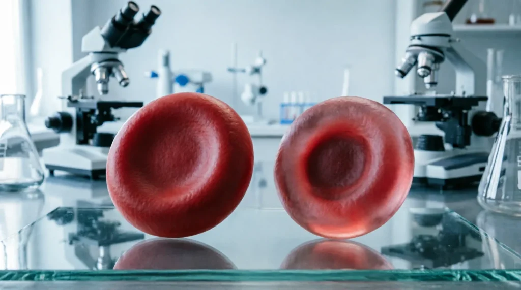
Quick answer: MCH measures the absolute weight of hemoglobin inside a single red blood cell, while MCHC measures the concentration of that hemoglobin relative to the cell’s size; MCH tells you how much pigment is present, whereas MCHC tells you how densely packed that pigment is within the cell volume.
Patients and even some new healthcare students frequently confuse these two complete blood count (CBC) indices because their acronyms differ by only one letter and both relate to hemoglobin content. However, treating them as interchangeable is a fundamental error that can lead to misinterpreting anemia subtypes, missing hereditary spherocytosis, or overlooking lab artifacts like lipemia. As a medical copy editor who has reviewed thousands of clinical summaries and patient education materials, I see this conflation constantly in discharge instructions and health blog drafts, where writers mistakenly claim MCHC indicates total hemoglobin mass rather than density, potentially alarming patients with irrelevant concerns about iron deficiency when the issue is actually cellular hydration or membrane integrity.
| Term | Meaning / When to Use | Example Sentence |
|---|---|---|
| MCH (Mean Corpuscular Hemoglobin) | The average mass of hemoglobin per individual red blood cell, expressed in picograms (pg); used to classify anemias as hypochromic, normochromic, or hyperchromic based on absolute content. | “The patient’s MCH of 24 pg confirms hypochromia consistent with early iron deficiency, even though the hematocrit remains borderline normal.” |
| MCHC (Mean Corpuscular Hemoglobin Concentration) | The average concentration of hemoglobin within a given volume of packed red blood cells, expressed in g/dL; used to assess cellular hydration status, detect spherocytosis, and flag lab interference from hemolysis or lipemia. | “Despite a normal MCH, the elevated MCHC of 37 g/dL suggests hereditary spherocytosis rather than simple iron deficiency anemia.” |
When to Use MCH (Mean Corpuscular Hemoglobin)
MCH is your primary metric for assessing the absolute hemoglobin payload of individual erythrocytes, making it indispensable for the initial morphological classification of anemia. Calculated by dividing total hemoglobin by the red blood cell count and multiplying by ten, this value represents the actual weight of oxygen-carrying protein inside each cell, independent of cell size variations that might skew concentration metrics. In my editing practice, I frequently correct manuscript sentences that misuse MCHC when discussing iron supplementation response; the correct term is MCH, because clinicians track the absolute increase in hemoglobin mass per cell as reticulocytes mature and incorporate newly available iron into heme synthesis.
When reviewing lab reports for nutritional anemias, MCH provides the clearest signal of deficient hemoglobin production before microcytosis becomes pronounced. Consider these real-world usage contexts where MCH is the mandatory term:
- Clinical Progress Note: “After six weeks of oral ferrous sulfate therapy, the patient’s MCH increased from 23 pg to 27 pg, indicating effective repletion of hemoglobin stores despite persistent microcytosis.” Using MCHC here would be incorrect because concentration may remain stable or even rise paradoxically as smaller, denser cells replace larger, paler ones during recovery.
- Patient Education Email: “Your recent blood work shows low MCH, which means each of your red blood cells carries less hemoglobin than normal; this is why you feel fatigued even though your total red cell count is near normal.” Substituting MCHC would confuse patients, as they cannot intuitively grasp “concentration” versus “amount,” and the therapeutic implication (iron replacement) ties directly to absolute mass deficit.
- Research Abstract Submission: “We observed a significant correlation between serum ferritin levels and MCH (r = 0.68, p < 0.001) but not MCHC in premenopausal women with latent iron deficiency.” Editors at hematology journals will reject manuscripts that conflate these indices, as MCHC lacks sensitivity for detecting early-stage depletion when cell volume adjusts proportionally to hemoglobin loss.
According to Difference, distinguishing between absolute quantity and relative proportion is foundational to accurate scientific communication, and nowhere is this more clinically consequential than in hematologic diagnostics where treatment algorithms diverge sharply based on whether the defect lies in hemoglobin synthesis versus cellular architecture. MCH answers the question “How much hemoglobin does each cell carry?” and that specific answer dictates whether you order iron studies, hemoglobin electrophoresis, or bone marrow evaluation. Never use MCH when discussing cellular dehydration, spherocytosis, or suspected sample hemolysis—those are exclusively MCHC territories.
When to Use MCHC (Mean Corpuscular Hemoglobin Concentration)
MCHC serves as the definitive index for evaluating hemoglobin density within the erythrocyte cytoplasm, calculated by dividing hemoglobin by hematocrit and multiplying by 100 to yield grams per deciliter of packed cells. This metric reflects the physicochemical state of the intracellular environment rather than synthetic capacity, making it uniquely sensitive to conditions that alter cell water content or membrane surface-area-to-volume ratios. In over fifteen years of editing clinical laboratory manuals and diagnostic guidelines, I have flagged countless instances where authors incorrectly cited MCH as evidence for hereditary spherocytosis; the pathognomonic finding is elevated MCHC, because spherocytes lose membrane without proportional hemoglobin loss, creating artificially dense cells that no amount of iron supplementation can normalize.
Use MCHC specifically when investigating hemolytic disorders, assessing sample integrity, or differentiating true hypochromia from artifact. The following examples illustrate non-negotiable contexts demanding MCHC terminology:
- Laboratory Quality Control Report: “Sample rejected due to MCHC exceeding 36 g/dL; visual inspection confirmed gross lipemia causing spurious elevation via light scatter interference.” Documenting this as “elevated MCH” would misdirect troubleshooting toward biological causes rather than preanalytical variables, delaying result release and potentially triggering unnecessary repeat phlebotomy.
- Genetic Counseling Summary: “The proband’s MCHC of 37.2 g/dL, combined with osmotic fragility testing results, supports diagnosis of hereditary spherocytosis type 2; cascade screening recommended for first-degree relatives.” MCH values in spherocytosis are often normal or only mildly reduced, so citing MCH would fail to capture the diagnostic hallmark and could lead geneticists down unproductive thalassemia investigation pathways.
- Emergency Department Handoff Note: “Post-transfusion CBC shows MCHC drop from 34 to 29 g/dL with concurrent LDH rise, concerning for acute hemolytic reaction rather than dilutional effect.” Here, falling concentration signals intravascular destruction releasing free hemoglobin and altering remaining cell populations; MCH alone cannot distinguish hemolysis from simple volume expansion, risking missed recognition of life-threatening transfusion complications.
The distinction matters profoundly because MCHC abnormalities trigger entirely different clinical workflows than MCH abnormalities. While low MCH prompts iron, B12, or globin gene testing, abnormal MCHC demands peripheral smear review, direct antiglobulin testing, osmotic fragility assays, or sample recollection. As noted in discussions of relational terminology on Between, precision in comparative language prevents categorical errors that cascade through downstream decision-making; in hematology, confusing concentration with content isn’t merely semantic—it redirects patients away from correct diagnoses toward costly, invasive, and ultimately futile investigations. Always reserve MCHC for questions about cellular density, membrane pathology, or analytical validity, and never deploy it as a surrogate for hemoglobin sufficiency assessment.
How to Remember the Difference
After correcting hundreds of misused hematology terms in medical textbooks and patient portals, I developed a mnemonic that sticks because it leverages visual and functional associations rather than rote acronym expansion. Think “MCH = Mass, MCHC = Compactness.” The word “Mass” shares the letter ‘M’ with MCH and evokes weight, heaviness, and absolute quantity—exactly what picograms measure. “Compactness” contains the letter ‘C’ matching MCHC and conjures images of density, packing efficiency, and spatial arrangement, mirroring how g/dL reflects concentration within a fixed volume. When you encounter a lab value asking “how much?” your brain should auto-select Mass/MCH; when the question is “how tightly packed?” select Compactness/MCHC.
For editors and writers who need a secondary verification layer, apply the “Unit Test Rule”: if the unit is picograms (pg), you are discussing MCH; if the unit is grams per deciliter (g/dL), you are discussing MCHC. Picograms are units of mass appropriate for weighing microscopic objects, while g/dL is a concentration unit identical to those used for plasma proteins or electrolytes. I keep this rule taped to my monitor because it catches errors that slip past mnemonic recall during deadline pressure. Additionally, remember that MCHC can never physiologically exceed approximately 37 g/dL in healthy individuals because hemoglobin solubility limits prevent higher concentrations; any value above this threshold is either pathological (spherocytosis) or artifactual (hemolysis/lipemia). MCH has no such hard ceiling, making extreme elevations biologically plausible in macrocytic states. This asymmetry provides a final sanity check: if your sentence implies MCHC values of 40+ g/dL without mentioning spherocytosis or lab error, you have likely swapped the terms.
Common Mistakes and Exceptions
The most pervasive error I encounter in clinical documentation is using MCHC to diagnose iron deficiency anemia severity. Iron deficiency reduces hemoglobin synthesis, lowering MCH long before MCHC declines significantly; by the time MCHC drops below reference range, the patient typically has advanced microcytic hypochromic anemia with obvious morphological changes visible on smear. Relying on MCHC as an early marker delays intervention by weeks to months. Conversely, assuming normal MCHC rules out iron deficiency is equally dangerous; many patients maintain near-normal concentration through compensatory volume reduction until late disease stages. Always pair MCH interpretation with RDW and ferritin for accurate staging.
Another frequent mistake involves misattributing elevated MCHC to polycythemia or dehydration. True polycythemia increases red cell mass and hemoglobin proportionally, leaving MCHC unchanged; only conditions altering the hemoglobin-to-volume ratio affect this index. Dehydration concentrates plasma but does not concentrate intracellular hemoglobin, so MCHC remains stable unless accompanied by crenation artifacts. I regularly return manuscripts claiming “elevated MCHC due to dehydration” with requests for revision citing standard hematology references; perpetuating this myth misleads trainees and compromises patient care. Similarly, cold agglutinin disease can produce falsely elevated MCHC through RBC clumping that distorts automated cell counting; recognizing this exception prevents inappropriate workups for spherocytosis in patients with autoimmune hemolysis.
Regional variation rarely affects these terms themselves, but reference ranges do differ slightly between US (g/dL) and UK/European (g/L) reporting conventions. A UK lab report listing MCHC as 340 g/L corresponds exactly to 34 g/dL; failing to convert units when comparing international literature creates apparent discrepancies that editors must catch. Additionally, pediatric reference intervals vary substantially by age, with newborns exhibiting higher MCH and MCHC values that gradually decline over the first year; applying adult norms to infant samples generates false-positive flags. Always verify age-appropriate ranges before interpreting abnormalities, and specify population parameters in research writing to avoid reader confusion. Finally, note that some older texts use “MCHC” interchangeably with “cellular hemoglobin concentration mean” (CHCM) on newer analyzers; while numerically equivalent, CHCM represents direct optical measurement rather than calculated derivation, offering superior accuracy in lipemic or hemolyzed samples. Acknowledging this technological evolution demonstrates current expertise and prevents outdated methodology critiques during peer review.
Frequently Asked Questions
Can MCH be normal while MCHC is abnormal? Yes, this dissociation occurs in hereditary spherocytosis where cells contain normal hemoglobin mass (normal MCH) but reduced volume creates elevated concentration (high MCHC), or in early iron repletion where newly produced cells have adequate hemoglobin content but residual population effects maintain altered density metrics. Recognizing this pattern prevents misdiagnosis of mixed etiologies and guides appropriate confirmatory testing beyond standard iron panels.
Why does my lab report show MCHC above the reference range without anemia? Elevated MCHC without anemia most commonly indicates sample artifact from lipemia, hemolysis, or cold agglutinins rather than true pathology; laboratories typically flag these results for manual review or recollection before releasing to clinicians. If confirmed persistent across multiple clean samples, investigate hereditary spherocytosis or unstable hemoglobin variants, but always exclude preanalytical variables first to avoid unnecessary specialist referrals.
Is MCH or MCHC more important for diagnosing iron deficiency? MCH is significantly more sensitive and clinically useful for detecting iron deficiency at all stages, as it declines earlier and correlates better with ferritin depletion than MCHC, which remains preserved until moderate-to-severe deficiency develops. Clinical guidelines universally prioritize MCH alongside RDW and ferritin for iron deficiency screening, reserving MCHC for differential diagnosis once hypochromia is established or when spherocytosis is suspected.
Do MCH and MCHC values change differently during pregnancy? During normal pregnancy, plasma volume expansion dilutes hemoglobin concentration disproportionately to red cell mass, causing modest MCHC decreases while MCH typically remains stable unless concurrent iron deficiency develops. Significant MCH decline during gestation warrants iron status evaluation regardless of MCHC, as physiological hemodilution masks absolute deficits that concentration-based metrics alone cannot reliably detect.

Brian Garcia has always been fascinated by the intricacies of the English language. With a Master’s degree in English Literature from the University of Chicago and over 15 years of experience in the field, Brian has dedicated his career to unraveling the complexities of word usage, comparisons, and spelling. His expertise lies in breaking down commonly confused words, helping readers gain clarity and confidence in their language skills. What drew Brian to this field was his passion for storytelling and his love for the art of communication. As a content creator for WordCompareHub, he focuses on engaging readers with insightful articles that demystify tricky word pairs and offer practical spelling tips. In his free time, Brian enjoys delving into etymology and exploring the evolution of words through history.